A Trio of Biomarkers: A Novel Multivariate Model to Detect DILI & Liver Damage
Posted on: September 27, 2021
A Trio of Biomarkers:
A Novel Multivariate Model to Detect DILI & Liver Damage
-Contributed by Abi Kasberg, PhD
While maybe underappreciated, Drug-induced liver injury (DILI) is a condition with significant impact on the medical world. DILI is the leading cause of acute liver failure and is frequently responsible for the termination and withdrawal of drugs from clinical trials and from the market. To overcome these clinical and economic challenges, there is a need for an expansion of available sensitive and specific biomarkers to more accurately determine and predict early stages of hepatotoxicity. A grand endeavor is underway to do just that. This endeavor is led by pharmaceutical and academic collaborations in coordination with third-parties, such as the Predictive Safety Testing Consortium (PTSC) of the Critical Path Institute (C-PATH), to serve as a third-party neutral voice. The goal of these collaborations is to identify and validate new safety DILI biomarkers for the FDA’s consideration. Pending FDA approval, this will open the opportunity for current hepatotoxicity guidelines to be expanded to include new clinical biomarker determinants of DILI and liver damage. Harnessing and leveraging the strength of more reliable, accurate, and specific DILI biomarkers will streamline early drug development processes and improve clarity for clinical evaluations of liver injury caused by xenobiotics.
Biomarkers traditionally used to detect DILI, both in drug development trials and in the clinic, are serum alanine aminotransferase (ALT), aspartate aminotransferase (AST), serum alkaline phosphatase (ALP), and total bilirubin concentration (TBL) (Church et al. 2017). Unfortunately, these commonly used biomarkers lack sensitivity and specificity for predicting early liver injury and are therefore not ideal for use during drug development studies (Church et al. 2017, Church et al. 2019). To circumvent these shortcomings, the Drug Safety Research and Development laboratory at Pfizer, Inc and collaborators evaluated the sensitivity and specificity of candidate biomarkers for the detection of liver injury (Llewellyn, et al. 2021). The objective of this study was to evaluate the diagnostic performance and ability of seven promising biomarkers to detect DILI and liver damage: total cytokeratin 18 (K18), caspase cleaved K18 (ccK18), macrophage colony-stimulating factor (MCSF), MCSF receptor (MCSFR), osteopontin (OPN), glutamate dehydrogenase (GLDH), and microRNA-122 (miR-122) (Llewellyn, et al. 2021). Results revealed that a panel of 3 biomarkers composed of K18, GLDH, and miR-122 are excellent at detecting DILI and predicting liver injury with more accuracy than any of the individual biomarkers tested, including ALT (Llewellyn, et al. 2021). Here, we review the individual background of each biomarker, K18, GLDH, and miR-122, and then explore potential explanations as to why the combined multivariate analysis using the three-biomarker panel is powerful at predicting liver injury.
K18, GLDH, and miR-122:
The Individuals
K18 is a type I intermediate filament (IF) protein that provides structural support to the cytoskeleton of epithelial cells. In the liver, K18 is predominantly found in hepatocytes and bile duct cholangiocytes (Ku et al. 2016, Llewellyn et al. 2021). K18 protects hepatocytes from apoptosis and necrosis, positioning it as a mechanistic biomarker of cell death (Ku et al. 2016). During necrosis, full-length K18 is passively released from cells following liver injury (Rupprechter et al. 2021). In contrast, during cell apoptosis, structural reorganizations of IFs drive caspase-mediated cleavage of full-length K18 into ccK18 (Caulin et al. 1998, Rupprechter et al. 2021). In this publication, total K18 and ccK18 are detected with ELISA kits M65® Epideath and M30 Apoptosense® (Llewellyn et al. 2021). Thus, measurements of total K18 levels and ccK18 levels in the blood provide insightful information regarding cell death processes during liver injury.
In this study, K18 displayed superior sensitivity and specificity over ALT, GLDH, and miR-122 for detecting liver damage in a cross-sectional cohort of patients (Llewellyn et al. 2021). In fact, K18 was suggested to function as a sufficient single biomarker for the assessment of liver injury (Llewellyn et al. 2021). Furthermore, it was shown that gene expression of KRT18, the gene that encodes K18, is elevated during liver apoptosis and necrosis, supporting that K18 plays an active role in liver damage (Llewellyn et al. 2021).
It is interesting to highlight that in the context of DILI, K18 is one of the three biomarkers used in the multivariate model for liver injury detection, whereas ccK18 is not (Llewellyn et al 2021). This suggests that measuring liver necrosis may be more relevant than apoptosis for the specific detection of DILI and acute liver damage. The ccK18 biomarker associates with liver injury, but shows reduced sensitivity and specificity compared to K18 for DILI and does not outperform ALT in distinguishing liver damage (Llewellyn et al. 2021). An explanation could be that the underlying mechanisms of DILI do not result in the activation of caspases and rapid release of ccK18, or perhaps the release of ccK18 from cells is inhibited during DILI.
GLDH is a mitochondrial enzyme that serves anabolic and catabolic functions. It is involved with urea production and contributes to the Krebs cycle (Plaitakis et al. 2017). Following mitochondrial swelling and damage during necrotic cell death, GLDH is released into the cytoplasm and further into circulation. Therefore, GLDH has been used as an early marker of mitochondrial damage and dysfunction (McGill et al. 2012, McGill et al. 2021). Serum GLDH levels are elevated during acetaminophen-induced acute liver failure, and GLDH has greater liver specificity than ALT to detect liver injury (Llewellyn et al. 2021, McGill et al. 2014). High serum levels of GLDH correlate with and are predictive of poor outcomes (McGill et al. 2014). In clinical studies, GLDH can differentiate liver injury from muscle injury, unlike ALT (Church et al. 2017). The half-life of GLDH is shorter than that of ALT, which suggests that GLDH more accurately reflects the status of ongoing liver injury (Church et al. 2017). Together, this makes GLDH a valuable mechanistic biomarker of mitochondrial damage that can be leveraged for the detection of DILI (Church et al. 2017, McGill et al. 2021).
Despite these advantages, GLDH has some drawbacks. For instance, GLDH may only be relevant to predict and detect DILI while considered in conjunction with other liver biomarkers (Roth et al. 2020). GLDH levels can increase in healthy individuals following treatment with drugs that are not associated with liver injury (Singhal et al. 2014). Fluctuating changes in GLDH levels have also been recorded following bile duct stone blockage and during circulatory abnormalities that drive liver hypoxia (Roth et al. 2020). GLUD1, the gene that expresses GLDH, is widely expressed with less liver specificity than KRT18 (Llewellyn et al. 2021). Therefore, GLDH is not a great stand-alone liver injury biomarker for DILI detection. However, GLDH is a valuable component of the multivariate model for liver injury detection, as determined by statistical random forest modeling (Lewellyn et al. 2021).
MiR-122 is a small noncoding microRNA that regulates post-transcriptional expression of gene targets, such as cationic amino acid transporter 1 (CAT-1) (Jopling et al. 2012). MiR-122 also functions to reduce plasma cholesterol levels through indirect regulation of the cholesterol biosynthesis pathway (Jopling et al. 2012). MiR-122 is strongly and specifically expressed in the liver. Following liver injury, miR-122 is released from hepatocytes undergoing necrosis and displays elevated levels in the circulation prior to the appearance of ALT (Rupprechter et al. 2020, Thulin et al. 2014, Wang et al. 2009). Similar to GLDH, miR-122 has a shorter half-life than ALT (Starkey Lewis et al. 2011). The presence of miR-122 levels in specific serum fractions has been shown to be useful for distinguishing alcoholic liver disease and liver inflammation from drug-induced liver necrosis (Bala et al. 2012, Thulin et al. 2017).
However, miR-122 levels are known to have significant inter- and intraindividual variability in healthy individuals (Church et al. 2019). Furthermore, it has been shown that miR-122 can be released from hepatocytes during subtoxic exposure to acetaminophen (Holman et al. 2016). This suggests that miR-122 can be actively released in the absence of hepatocyte death, likely in response to stress (Church et al. 2019). Nevertheless, miR-122 is an early and specific liver injury biomarker that can be used to investigate mechanisms of liver pathologies. MiR-122 is also a vital component of the three biomarker-based multivariate model of liver damage detection during DILI.
K18, GLDH, and miR-122:
A Multivariate Model for Liver Injury Detection
When considered individually, the liver biomarkers K18, GLDH, and miR-122 provide mechanistic insights into liver damage and hepatocyte death, but each biomarker has its own vulnerabilities. When analyzed together in a multivariate model, however, these biomarkers provide sensitive and liver-specific data for the accurate prediction and detection of liver injury that outperforms each individual biomarker alone (Llewellyn et al. 2021). The impressive ability of this three-biomarker panel to accurately predict and detect DILI could in part be explained by the common characteristics shared by all three biomarkers (hepatocyte expression and necrosis). These shared attributes are complemented by diverse biomarker activities (cellular localization, function, and specificity).
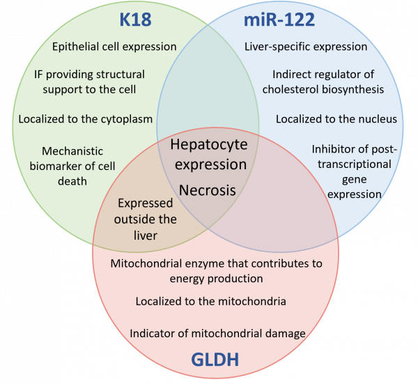
Figure 1: Venn Diagram of common and distinct attributes of DILI biomarkers K18, GLDH, and miR-122. K18, total cytokeratin 18; GLDH, glutamate dehydrogenase; miR-122, microRNA-122; IF, intermediate filament
There are two common attributes that are shared by these three biomarkers: elevated hepatocyte expression and the exit pathway from the cell via necrosis (Fig. 1). The fact that each biomarker in the three-biomarker panel has these two properties suggests that these characteristics are important for the sensitive and specific detection of liver injury and DILI potential. Cells that are directly responding to the trauma of liver injury, namely hepatocytes, are likely to be the first cells to immediately react with increased biomarker production. The second attribute, necrosis, is responsible for the early and rapid detectability of each biomarker in the blood. As hepatocytes necrotize, they are not only dying in response to hepatotoxicity, but they are also releasing biomarkers into the circulation. This is perhaps a critical attribute due to the nature of sample collection. Subject samples must be collected in a manner that is accessible and minimally invasive. This makes bodily fluids such as blood a popular sample option to collect. Hence, it is critical that excellent DILI biomarkers be detectable in serum or plasma. There are additional explanations as to why necrosis may be the primary mechanism of biomarker release from the cell during DILI instead of other pathways. Rapid mitochondrial decline is linked to hepatocyte necrosis (Antoine et al. 2013). GLDH is a marker of mitochondrial dysfunction, and hence the presence of GLDH on the three-biomarker panel could explain why necrosis, and not apoptosis, appears to be a critical contributor to DILI pathogenesis (Antoine et al. 2013). The three biomarkers that make up the multivariate model for liver injury detection share increased expression in hepatocytes and are all released following necrosis, suggesting that these two attributes are indispensable for DILI biomarker efficacy.
There are several key differences to highlight when considering the multivariate impact of GLDH, K18, and miR-122 biomarkers. The first is the subcellular localization of each biomarker within hepatocytes prior to necrosis. GLDH is localized to the mitochondria, K18 is in the cytoplasm, and miR-122 is localized to the nucleus of hepatocytes (Fig. 1). Secondly, they have functions independent from each other within the cell. GLDH is a mitochondrial enzyme involved with energy production, K18 is a structural intermediate filament protein, and miR-122 is a regulator of post-transcriptional expression. Perhaps the diversity of cellular function and localization casts a wide net of detectability, enabling a greater range and potential of biomarker exposure in the blood. The three-biomarker panel detects the downstream effects of dying cells that have undergone mitochondrial crises, their cellular structural support is decaying, and post-transcriptional gene regulation pathways are disrupted. This multivariate detection strategy enables a multifaceted snapshot of liver health as a strategic means to detect early stages of DILI. In contrast, the detection of a single biomarker with associated individual cellular attributes may go undetected or sound an unnecessary alarm.
It is illuminating to note what is absent from the multivariate panel of three DILI biomarkers; what did not make the statistical cut. Candidate biomarkers that are released from apoptotic cells (ccK18), macrophages (MCSFR1, OPN), lymphocytes (OPN), or biomarkers required for the maturation of macrophages (MCSF) were not selected by random forest modeling for their ability to predict ALT levels and the associated liver damage during acetaminophen overdose (Llewellyn et al. 2021). This suggests that immune response biomarkers may not be as sensitive or are possibly delayed in their ability to predict liver injury compared to biomarkers that are directly expressed in hepatocytes undergoing necrosis. As discussed previously, apoptosis and the biomarker ccK18 do not appear to be the best indicators of DILI when compared to the mechanisms of necrosis and K18.
As expected, increasing the number of measurable parameters increases the statistical strength of the multivariate model. This is supported through analysis of a seven biomarker-based model (K18, GLDH, miR-122, ccK18, MCSF, MCSFR, and OPN) compared to the three biomarker-based model (K18, GLDH, miR-122). However, the seven biomarker-based model exhibited similar DILI predictive value and had only slightly greater specificity for identifying liver damage compared to the three biomarker-based model (Llewellyn et al. 2021). The practical limitations to running a seven-biomarker panel may not outweigh the modest benefits. In conclusion, the three-biomarker multivariate panel that contains K18, GLDH, and miR-122 was found to be an accurate and sensitive indicator of liver injury (Llewellyn et al. 2021). This multivariate model could be instrumental in improving the specificity and sensitivity of DILI detection for use in drug development studies and clinical trials.
Further Reading
Antoine DJ, Dear JW, Lewis PS, Platt V, Coyle J, Masson M, Thanacoody RH, Gray AJ, Webb DJ, Moggs JG, Bateman DN, Goldring CE, Park BK. Mechanistic biomarkers provide early and sensitive detection of acetaminophen-induced acute liver injury at first presentation to hospital. Hepatology. 2013 Aug;58(2):777-87. doi: 10.1002/hep.26294. Epub 2013 Jul 2. PMID: 23390034; PMCID: PMC3842113.
Bala S, Petrasek J, Mundkur S, Catalano D, Levin I, Ward J, Alao H, Kodys K, Szabo G. Circulating microRNAs in exosomes indicate hepatocyte injury and inflammation in alcoholic, drug-induced, and inflammatory liver diseases. Hepatology. 2012 Nov;56(5):1946-57. doi: 10.1002/hep.25873. Epub 2012 Jul 26. PMID: 22684891; PMCID: PMC3486954.
Caulín C, Salvesen GS, Oshima RG. Caspase cleavage of keratin 18 and reorganization of intermediate filaments during epithelial cell apoptosis. J Cell Biol. 1997 Sep 22;138(6):1379-94. doi: 10.1083/jcb.138.6.1379. PMID: 9298992; PMCID: PMC2132555.
Church RJ, Watkins PB. The transformation in biomarker detection and management of drug-induced liver injury. Liver Int. 2017 Nov;37(11):1582-1590. doi: 10.1111/liv.13441. Epub 2017 May 8. PMID: 28386997; PMCID: PMC5632128.
Church RJ, Kullak-Ublick GA, Aubrecht J, Bonkovsky HL, Chalasani N, Fontana RJ, Goepfert JC, Hackman F, King NMP, Kirby S, Kirby P, Marcinak J, Ormarsdottir S, Schomaker SJ, Schuppe-Koistinen I, Wolenski F, Arber N, Merz M, Sauer JM, Andrade RJ, van Bömmel F, Poynard T, Watkins PB. Candidate biomarkers for the diagnosis and prognosis of drug-induced liver injury: An international collaborative effort. Hepatology. 2019 Feb;69(2):760-773. doi: 10.1002/hep.29802. Epub 2018 Jun 27. PMID: 29357190; PMCID: PMC6054900.
Holman NS, Mosedale M, Wolf KK, LeCluyse EL, Watkins PB. Subtoxic Alterations in Hepatocyte-Derived Exosomes: An Early Step in Drug-Induced Liver Injury? Toxicol Sci. 2016 Jun;151(2):365-75. doi: 10.1093/toxsci/kfw047. Epub 2016 Mar 8. PMID: 26962055; PMCID: PMC4880137.
Ku NO, Strnad P, Bantel H, Omary MB. Keratins: Biomarkers and modulators of apoptotic and necrotic cell death in the liver. Hepatology. 2016 Sep;64(3):966-76. doi: 10.1002/hep.28493. Epub 2016 Apr 4. PMID: 26853542; PMCID: PMC4977204.
Llewellyn HP, Vaidya VS, Wang Z, Peng Q, Hyde C, Potter D, Wang J, Zong Q, Arat S, Martin M, Masek-Hammerman K, Warner R, Johnson K, Kullak-Ublick GA, Aithal GP, Dear JW, Ramaiah SK. Evaluating the Sensitivity and Specificity of Promising Circulating Biomarkers to Diagnose Liver Injury in Humans. Toxicol Sci. 2021 Apr 27;181(1):23-34. doi: 10.1093/toxsci/kfab003. PMID: 33483742.
McGill MR, Sharpe MR, Williams CD, Taha M, Curry SC, Jaeschke H. The mechanism underlying acetaminophen-induced hepatotoxicity in humans and mice involves mitochondrial damage and nuclear DNA fragmentation. J Clin Invest. 2012 Apr;122(4):1574-83. doi: 10.1172/JCI59755. Epub 2012 Mar 1. PMID: 22378043; PMCID: PMC3314460.
McGill MR, Staggs VS, Sharpe MR, Lee WM, Jaeschke H; Acute Liver Failure Study Group. Serum mitochondrial biomarkers and damage-associated molecular patterns are higher in acetaminophen overdose patients with poor outcome. Hepatology. 2014 Oct;60(4):1336-45. doi: 10.1002/hep.27265. Epub 2014 Aug 25. PMID: 24923598; PMCID: PMC4174728.
McGill MR, Jaeschke H. Biomarkers of mitotoxicity after acute liver injury: Further insights into the interpretation of glutamate dehydrogenase. J Clin Transl Res. 2021 Feb 25;7(1):61-65. PMID: 34027202; PMCID: PMC8132186.
Plaitakis A, Kalef-Ezra E, Kotzamani D, Zaganas I, Spanaki C. The Glutamate Dehydrogenase Pathway and Its Roles in Cell and Tissue Biology in Health and Disease. Biology (Basel). 2017 Feb 8;6(1):11. doi: 10.3390/biology6010011. PMID: 28208702; PMCID: PMC5372004.
Roth SE, Avigan MI, Bourdet D, Brott D, Church R, Dash A, Keller D, Sherratt P, Watkins PB, Westcott-Baker L, Lentini S, Merz M, Ramaiah L, Ramaiah SK, Stanley AM, Marcinak J. Next-Generation DILI Biomarkers: Prioritization of Biomarkers for Qualification and Best Practices for Biospecimen Collection in Drug Development. Clin Pharmacol Ther. 2020 Feb;107(2):333-346. doi: 10.1002/cpt.1571. Epub 2019 Sep 14. PMID: 31314926; PMCID: PMC7006882.
Rupprechter SAE, Sloan DJ, Oosthuyzen W, Bachmann TT, Hill AT, Dhaliwal K, Templeton K, Matovu J, Sekaggya-Wiltshire C, Dear JW. MicroRNA-122 and cytokeratin-18 have potential as a biomarkers of drug-induced liver injury in European and African patients on treatment for mycobacterial infection. Br J Clin Pharmacol. 2021 Aug;87(8):3206-3217. doi: 10.1111/bcp.14736. Epub 2021 Jan 26. PMID: 33432705.
Singhal R, Harrill AH, Menguy-Vacheron F, Jayyosi Z, Benzerdjeb H, Watkins PB. Benign elevations in serum aminotransferases and biomarkers of hepatotoxicity in healthy volunteers treated with cholestyramine. BMC Pharmacol Toxicol. 2014 Aug 3;15:42. doi: 10.1186/2050-6511-15-42. PMID: 25086653; PMCID: PMC4130124.
Starkey Lewis PJ, Dear J, Platt V, Simpson KJ, Craig DG, Antoine DJ, French NS, Dhaun N, Webb DJ, Costello EM, Neoptolemos JP, Moggs J, Goldring CE, Park BK. Circulating microRNAs as potential markers of human drug-induced liver injury. Hepatology. 2011 Nov;54(5):1767-76. doi: 10.1002/hep.24538. PMID: 22045675.
Thulin P, Nordahl G, Gry M, Yimer G, Aklillu E, Makonnen E, Aderaye G, Lindquist L, Mattsson CM, Ekblom B, Antoine DJ, Park BK, Linder S, Harrill AH, Watkins PB, Glinghammar B, Schuppe-Koistinen I. Keratin-18 and microRNA-122 complement alanine aminotransferase as novel safety biomarkers for drug-induced liver injury in two human cohorts. Liver Int. 2014 Mar;34(3):367-78. doi: 10.1111/liv.12322. Epub 2013 Oct 14. PMID: 24118944.
Thulin P, Hornby RJ, Auli M, Nordahl G, Antoine DJ, Starkey Lewis P, Goldring CE, Park BK, Prats N, Glinghammar B, Schuppe-Koistinen I. A longitudinal assessment of miR-122 and GLDH as biomarkers of drug-induced liver injury in the rat. Biomarkers. 2017 Jul;22(5):461-469. doi: 10.1080/1354750X.2016.1269131. Epub 2016 Dec 15. PMID: 27978773.
Wang K, Zhang S, Marzolf B, Troisch P, Brightman A, Hu Z, Hood LE, Galas DJ. Circulating microRNAs, potential biomarkers for drug-induced liver injury. Proc Natl Acad Sci U S A. 2009 Mar 17;106(11):4402-7. doi: 10.1073/pnas.0813371106. Epub 2009 Feb 25. PMID: 19246379; PMCID: PMC2657429.
ML-00-00860 Rev01