
Diagnostics, Biomarkers, Instruments & Scientific Expertise
DiaPharma provides specialty research and clinical assay kits and instrumentation with personalized scientific expertise and customer support.
Products
DiaPharma specializes in assays and mechanistic biomarkers for bleeding and clotting disorders, hemostasis analyzers, liver disease, drug-induced liver and kidney injury, and anti-cancer drug development.
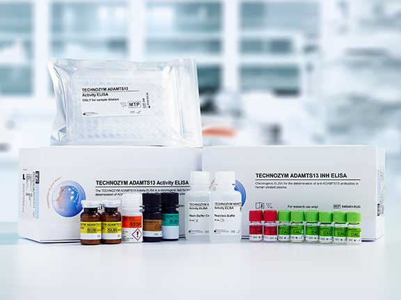
Hemostasis
TTP, Hemophilia, Thrombophilia, and more
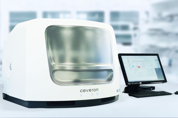
Instruments
Primary and Secondary Hemostasis
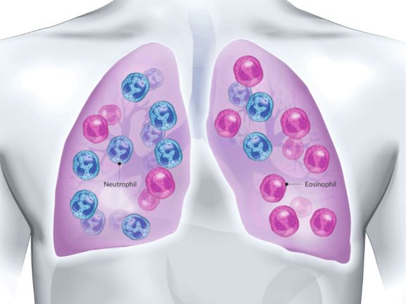
Hematology
Flow Cytometry Antibodies
Liver Disease
MASH, ALD, DILI Biomarker Assays
Fibrosis
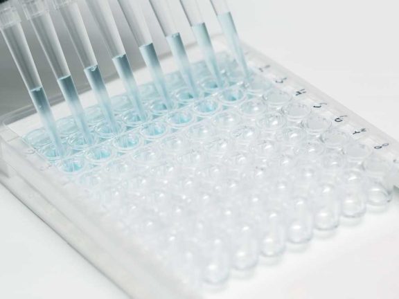
Assay Kits and Reagents
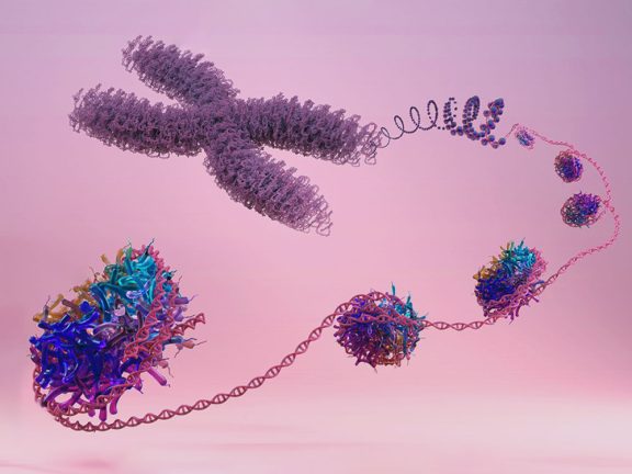
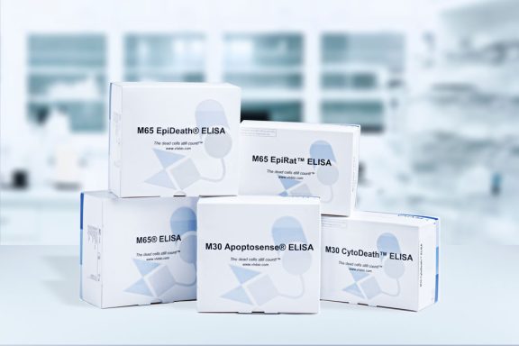
NETosis Research
Nucleosome ELISAs
Additional Biomarkers
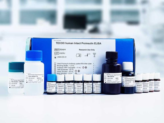
Biomarker Assay Kits

About Us
DiaPharma provides strong personal support, technical competence and experience to ensure customer expectations will be met or exceeded. Our strength is grounded in both science and staff. For over 25 years, DiaPharma has been proud to give each of our customers a truly personalized experience — a focused, dedicated resource that understands our customers’ needs and has the scientific expertise to help customers make the best product choices.
Stay Connected
Sign up for our newsletter
Enter your email below to stay up to date with industry news and get information about our products and events!
Differential Discussions Guest Series: The Use of Chromogenic Factor X Assays for Monitoring Patients on Warfarin Therapy
Posted: April 11, 2025
David L. McGlasson, MS, MLS(ASCP) A recent episode of “Differential Discussions” digs into the utility…
Through the Lens of CK18: Reflections on the AASLD ETC 2025: metALD and ALD
Posted: March 31, 2025
— Contributed by Olivia Stricker, PhD and Jessica Tuohy, PhD The 2025 AASLD Emerging Topics…