Heparin is a naturally occurring anticoagulant that prevents the formation and extension of blood clots. Heparin does not break down clots that have already formed (unlike tissue plasminogen activator) but allows fibrinolysis to work normally to break down clots.
Heparin is a widely used blood thinner used to prevent and treat deep vein thrombosis, pulmonary embolism, and myocardial infarction and is on the World Health Organization’s List of Essential Medicines.
Unfractionated heparin is heparin pharmaceutical that has not been fractionated to sequester the fraction of molecules with low molecular weight. Low molecular weight heparin has undergone fractionation for the purpose of making its pharmacodynamics more predictable.
Current clinical laboratory assays for heparin rely on an indirect measurement of the effect of the drug, rather than on a direct measure of its chemical presence. These include activated partial thromboplastin time (APTT) and antifactor Xa activity. Diapharma’s heparin kits are for the chromogenic determination of UF and LMW heparin in human plasma, and measure the ability of heparin to catalyze the inhibition of FXa by antithrombin.
From Ancient Greek ἧπαρ (hêpar) “liver”.
Introduction
Heparin preparations extracted from animals have been used clinically for over half a century as a potent anticoagulant therapeutic for the treatment and prevention of thrombotic disease. Bleeding and heparin-induced thrombocytopenia are the main adverse reactions associated with heparin therapy. These risks can be minimized by appropriate patient management and by laboratory monitoring using specific chromogenic anti-factor Xa assays. The clinical indications for these assays are reviewed together with a basic introduction of the clinical pharmacology of heparin.
Heparin is a naturally occurring, highly sulfated polysaccharide, characterized by a wide molecular weight range and powerful anticoagulant properties. Since its discovery by McLean in 1916, heparin has become a widely used anticoagulant for the treatment and prevention of thrombotic diseases and for maintaining blood fluidity in extracorporeal devices. Material for clinical use is derived from porcine and bovine tissue and is prepared either as unfractionated (UF) heparin or as depolymerized low molecular weight (LMW) heparin.
The main complication with heparin therapy is that it occasionally causes life-threatening bleeding. Laboratory monitoring with adjustments of doseregimens is one of the options available which may improve the antithrombotic efficacy of heparin and reduce the risk of hemorrhage. However, the ideal heparin test and its clinical relevance is still a controversial topic.
| Observed Reaction | Incidence |
|---|---|
| Hemorrhage | 1-33% |
| Thrombocytopenia | 1-3% |
| Hypertransaminasemia | up to 93% |
| Osteoporosis (long term use) | no data |
| Allergic reactions | no data |
| Cutaneous reactions | no data |
| Drug interactions | no data |
UF heparin activity is usually estimated by a conventional clotting test such as the activated partial thromboplastin time (APTT), the thrombin clotting time (TCT), or the activated clotting time (ACT). These assays are non-specific and are termed ‘global assays’ as they reflect the ability of heparin to interfere with several steps in the coagulation cascade. APTT is today the most widely-used test for heparin, mainly because it is relatively simple and allows for automation. However, when a test such as the APTT is prolonged beyond the normal range or does not reflect the anticipated effect of heparin, a more specific assay is required.
The specific assays measure the effect of heparin-accelerated antithrombin on a single coagulation enzyme, either factor Xa or thrombin (factor IIa). The enzyme is determined by its clotting activity (chronometrically) or by its activity against chromogenic peptide substrates (amidolytically). A major advantage of the specific assays is that they isolate the biological activity of heparin to one reaction, thus minimizing the interference from other variables. The anti-factor Xa assay is the only practical way of measuring LMW heparins.
Heparin data
| Trade names: | Heparin sodium, Liqueminsodium, Lipo-Hepin, Panheprin Calciparin etc. |
|---|---|
| Biosynthesis: | Mast cells |
| Specific activity: | 150-190 IU/mg |
| AT- high affinity chains: | 30-35% |
| Molecular weight: | 5,000 to 30,000 daltons |
| Structure: | Sulfated polysaccharide of 12 to over 100 saccharides Half-life: 1 to 3 hours (dose-dependent ) |
| Bioavailability: | ~ 30% |
| Removal pathway: | Cellular (saturable mechanism), renal (insaturable mechanism) |
| Function: | Accelerates primarily the inhibition of thrombin and factor Xa |
LMW Heparin data
| Trade names: | Fraxiparin, Enoxaparin, Fragmin, Sandoparin, Logiparin, Lovenox etc. |
|---|---|
| Method of preparation: | nitrous acid digestion, heparinase digestion, peroxidative cleavage, Β-elimination of heparin ester, benzylation followed by alkaine hydrolysis, nitrous acid depolymerrisation |
| Specific activity: | 80-120 IU/mg (anti-Xa assay) |
| AT- high affinity chains: | <30% |
| Mean molecular weight: | 4,000 to 6,500 daltons |
| Structure: | Sulfated polysaccharide of 4 to 40 saccharides Half-life: 4 hours (dose-dependent) |
| Bioavailability: | ~ 90% |
| Removal pathway: | Renal |
| Function: | Accelerates primarily the inhibition of factor Xa |
Biochemistry of Heparin
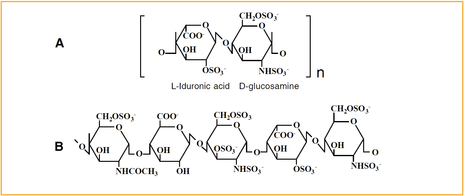
Heparin is a heterogenous mixture of polysaccharides, which chains are made up of alternating 1 to 4 linked, sulfated monosaccharide residues of L-iduronic acid and D-glucosamine. [A] is the most frequent type of disaccharide unit, representing up to 90% of the structure of beef-lung heparin, and up to 70% of pig-mucosa heparin. [B] is the unique pentasaccharide binding site for antithrombin which occurs in about one-third of the heparin chains.
Structure and biological role
Heparin is a sulfated glycosaminoglycan (GAG) mixture, which consists of unbranched polysaccharide chains, composed of 15 to 100 alternating monosaccharide units of L-iduronic acid and Dglucosamine. It has been found in mast cells in a large number of mammalian and nonmammalian vertebrates and is located mainly in tissues/organs that are in direct contact with the environment (i.e. lung, skin and intestine).
The extravascular location of heparin and the failure to detect it in blood have suggested that heparin does not normally have a role in regulating blood coagulation. However, heparan sulfate, a heparin-related GAG located on endothelial cells which line the blood vessel wall, have been shown to have anticoagulant activity. This fact could explain the ability of heparin to interfere with blood coagulation.
Anticoagulant activity
The basis for heparin’s (and heparan sulfate’s) anticoagulant activity in plasma is that it binds to antithrombin, the major inhibitor of the coagulation cascade in plasma. Binding induces a conformational change in the antithrombin molecule, which greatly accelerates the antithrombin inhibition of several serine proteases, including factors IXa, Xa, XIa, XIIa, kallikrein and thrombin.
The result is a stable 1:1 proteaseinhibitor complex, which is rapidly removed from the circulation and catabolized. The heparin-accelerated inhibition of thrombin and factor Xa constitutes the major portion of heparin’s anticoagulant effect in vitro, and possibly also the antithrombotic effect in vivo.
Catalytic mechanism
Heparin accelerates the inactivation of thrombin, and presumably also factors IXa and XIa, by serving as a template to which both antithrombin and the protease bind to form a ternary complex.
The accelerating function of heparin depends on the presence of a unique antithrombin-binding pentasaccharide sequence in a heparin GAG chain. Binding to this pentasaccharide induces a conformational change in the antithrombin molecule, which facilitates the reaction with the target protease.
Thrombin initially binds to the antithrombin-heparin complex in a non-specific fashion to any site along the GAG chain, then it slides along the surface until it encounters the inhibitor. It has been found that this sliding mechanism for thrombin requires a GAG chain of at least 18 monosaccharide units (Mw > 5,400 Da). Surprisingly, the sliding mechanism is not required for the inhibition of factors Xa, XIIa or kallikrein. Instead, the inhibition works predominantly through the conformational change of antithrombin bound to heparin.
The accelerated inactivation of these enzymes can therefore be achieved by GAG chains as small as the unique pentasaccharide sequence (Mw = 1,756 Da).
Another important feature of heparin is that its affinity for the antithrombin-protease complex is much lower than that of unreacted antithrombin. Heparin will therefore dissociate from the complex once its job is done, being free to catalyze further antithrombin reactions.
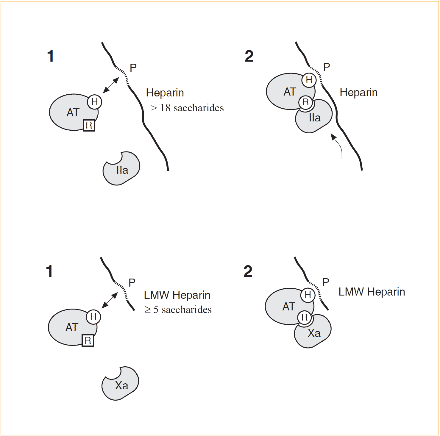
Model describing how heparin catalyzes the antithrombin-protease reaction
[H] symbolizes the heparin binding site and [R] is the reactive site in antithrombin, normally in an unfavorable conformation for protease inhibition. [P] is the unique antithrombin binding sequence of heparin. Binding to this sequence induces a conformational change in antithrombin ,which facilitates its reaction with its target proteases.
Top: The effect of heparin on the reaction between antithrombin and thrombin (IIa) involves binding both the enzyme and the inhibitor to the heparin chain, which thus needs to be of a certain length (≥18 monosaccharides). Thrombin binds to heparin in a non-specific manner (through positive surface charges) and ‘slides’ along the chain until it encounters the bound antithrombin.
Bottom: Inactivation of factor Xa does not require a ternary complex formation (i.e. the sliding mechanism is not required) and is achieved solely through heparin binding to antithrombin. Heparin’s affinity to the antithrombin-protease complex is much lower than that of free antithrombin and therefore heparin dissociates and binds to unreacted antithrombin, thus being able to catalyze further antithrombin reactions
Molecular weight aspects
Two forms of heparin are used clinically: unfractionated (UF) heparin with an average molecular weight of 15,000 (range 15 to 100 monosaccaharides) and low molecular weight (LMW) heparin with molecular weights between 4,000 to 6,500 (range 4 to 40 monosaccaharides). The reduction in molecular weight causes a marked change in the heparin activity. LMW heparin acts primarily on FXa, whereas UF heparin is an efficient catalyst for inhibition of both thrombin and factor Xa.
Unfractionated heparin
Two distinct fractions can be obtained from UF heparin by using affinity chromatography with immobilized antithrombin. The fraction that accounts for roughly 30% of the starting material, and nearly all the anticoagulant activity, is known as high-affinity heparin. The other fraction, which represents the majority of heparin chains, is the lowaffinity heparin, with virtually no anticoagulant activity. The different anticoagulant activities of these two fractions are the result of the unique antithrombin-binding pentasaccharide sequence, which is absent in low-affinity heparin chains.
The anticoagulant activity or potency of UF heparin is expressed relative to the 4th international standard. UF heparin preparations have specific activities of 150-190 IU/mg.
LMW heparin
The term low molecular weight (LMW) heparin refers to a heparin preparation obtained by fractionation of natural low-molecular weight material in UF heparin or by depolymerization of UF heparin.
Reduction in chain length of heparin reduces its affinity to plasma proteins, vascular matrix proteins, endothelial cells, marcrophages and platelets. As a result, LMW heparins have greater bioavailability, a longer plasma half-life, a more predictable therapeutic response to fixed doses and reduced plateletassociated side-effects.
A characteristic feature of LMW heparins is that they have less ability to enhance thrombin inibition than to potentiate factor Xa inhibition compared to UF heparin. The difference may be described in terms of an activity ratio such as the anti-factor Xa/ anti-factor IIa ratio. For UF heparins the ratio is 1:1 whilst for LMW Heparin the ratio is 1:2-1:4.
The anticoagulant activity or potency of LMW heparin is expressed relative to the 1st international standard for LMW heparin. The specific activities among LMW Heparins varies between 80-120 antiXa U/mg and between 35-45 anti-IIa U/mg.
Clinical Aspects of Heparin
Clinical use of heparin
Established uses for heparin include the treatment and prevention of various thrombotic disorders. Heparins are also used as an anticoagulant in extracorporeal circulations or in dialysis devices. Therapeutic objective The basic aim of using heparin preparations clinically is to reduce, delay or prevent the presence of thrombin. When heparin is used for prophylaxis (low-dose regimens), thrombin generation is mainly prevented. In the case of acute thrombosis, heparin is used for neutralizing thrombin that has already been formed, and for preventing further thrombin generation (high-dose regimens).
Therapeutic objective
The basic aim of using heparin preparations clinically is to reduce, delay or prevent the presence of thrombin. When heparin is used for prophylaxis (low-dose regimens), thrombin generation is mainly prevented. In the case of acute thrombosis, heparin is used for neutralizing thrombin that has already been formed, and for preventing further thrombin generation (high-dose regimens).
Mode of administration
Administration of heparin is performed by intravenous or subcutaneous routes, as intermittent injections or continuous infusion. The effect is immediate when given intravenously, whereas the action of subcutaneous heparin occurs within 20 to 60 minutes. The systemic absorption of heparin by oral or nasal administration is neglible.
Pharmacokinetics
The elimination rate of heparin from the blood is dose dependent. With low doses the clearance of UF heparin appears to rely on a saturable mechanism caused mainly by endothelial cell-uptake. At high doses a non-saturable mechanism predominates due to renal filtration.
LMW heparins are cleared mainly by renal filtration, probably due to its lower affinity to endothelial cells. As a result, LMW heparins have a two to four times longer half-life as compared to UF heparin at therapeutic doses, and ~90% bioavailability following subcutaneous injection as compared to only ~30% for UF heparins. This enables LMW heparins to be administered as a single daily injection.
Treatment of venous thromboembolism
Heparin has a long clinical history as the principle therapeutics in acute therapy for both deep vein thrombosis (DVT) and pulmonary embolism. Several studies have confirmed heparin’s role in the treatment of thrombosis.
After a 5 or 10-day course of heparin, treatment with the anticoagulant warfarin is usually started and then continued for several months.
Heparin resistance
Patients with thromboembolism that require more than 35,000 U/24 h to achieve the therapeutic range are classified as ‘heparin resistant’. There are a number of possible causes, such as increased heparin clearance, increased levels of procoagulants, reduced antithrombin levels, and increased levels of heparin binding proteins (e.g. platelet factor 4 and histidine rich glycoprotein).
Prophylaxis of DVT
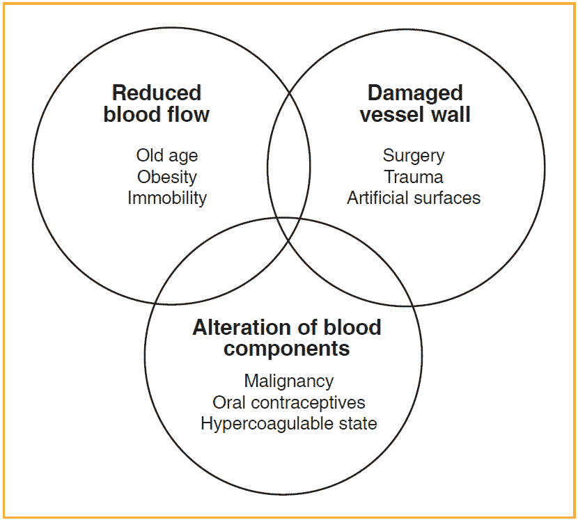
A reduced blood flow, the alteration of blood components and abnormalities of blood vessels are predisposing risk factors believed to result in thrombosis.
High-risk groups for the development of DVT include patients with acute myocardial infarction and patients who have undergone major surgery such as abdominal and orthopedic operations. In general surgical procedures the rate of DVT formation may be as high as 28% and even up to 50% after open prostatectomy or hip fracture.
Numerous clinical trials have demonstrated the efficacy of heparin therapy in reducing the incidence of pulmonary embolism and DVT as well as the long term complications after major surgery. The effectiveness of several commercial LMW heparins have also been investigated, most of which suggest a relative superiority (i.e. increased convenience) when compared to UF heparin. Although they are currently more expensive, they have proven to be cost effective for prophylaxis of DVT.
Extracorporeal circulation
Exposure of blood to large artificial surfaces (e.g. haemodialysis, cardiopulmonary bypass) activates coagulation. This may lead to thrombus formation and impaired function or occlusion of medical devices. The normal procedure is to control coagulation by administering UF heparin, although recent developments, which may promise fewer bleeding problems, include the use of LMW heparins or the concept of using heparin-coated membranes.
Complications
Hemorrhage
Hemorrhage is the main complication associated with heparin therapy, particularly when full dose heparin is injected intravenously. Major bleeding has been reported to occur in 1 to 33% of patients receiving various forms of heparin therapy. The risk is greater in the elderly, in patients with hypertension after trauma or surgery, and in patients with additional hemostatic abnormalities.
In summary, there are four variables reported to influence the risk of bleeding: the dose, the patient’s anticoagulant response, the mode of administration and specific patient-related characteristics.
Thrombocytopenia
Heparin-induced thrombocytopenia is another adverse effect with a reported incidence of 1-3%. The effect is usually moderate and is reversible once heparin administration is discontinued.
Occasionally a more severe heparin-induced thrombocytopenia (platelet count less than 50,000/µl) may occur, causing acute arterial thrombosis (‘white clot syndrome’).
Other toxicities and drug interactions
Following long-term heparin therapy, the development of osteoporosis (bone loss) can occur, with vertebral fractures as the predominant clinical sign. Most cases reported are in connection with pregnancy. Hypertransaminasemia has been observed in as many as 93% of subjects receiving heparin. Cases of skin necrosis have been observed with both UF heparin and LMW heparin. The precise cause of these heparin-induced reactions is at present unclear.
Heparin has been shown to cause a prehaemorrhagic tendency in patients undergoing aspirin (salicylates) therapy. Of particular note is the fact that heparin may be inhibited by concurrent intravenous nitroglycerin infusion during the treatment of patients with unstable angina or in the acute postmyocardial infarction period.
Laboratory monitoring of heparin
The clinical relevance
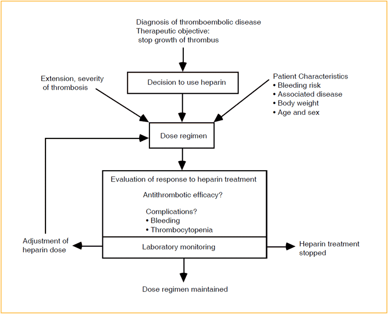
The purpose of monitoring heparin therapy is primarily to minimize the risk of haemorrhage from overdosage and to optimize the antithrombotic effect by appropriate dose adjustments. However, relatively few studies have been performed that clearly evaluate the usefulness of laboratory monitoring.
The main reason is the complex pharmacokinetics of heparin and the relatively weak correlation between the antithrombotic effect (in vivo) and anticoagulant activity (in vitro). Nevertheless, it is generally accepted that high-dose intravenous therapy with UF heparin should be monitored because of the danger of haemorrhage. For LMW heparins there is currently no definitive recommendation in favor of monitoring although, considering the comparable hemorrhagic risk of LMW heparins to UF heparins, as well as the risk of undertreatment, it may be useful to test the anti-Xa activity at least once at the beginning of treatment.
Low doses of UF heparin or LMW heparin, given subcutaneously for prophylaxis of DVT seldom require monitoring, although it may be useful to document the anti-Xa level in the case of unexpected hemorrhagic episodes.
Individual response to heparin
The risk of bleeding depends not only on the dose but also on the individual response. A large number of variables influence the antithrombotic and anticoagulant effect of heparin, including sex, age, weight, drug interactions, associated disease, extent of fibrin, vascular surfaces and the levels of various heparinbinding proteins. Elevated levels of heparin-binding proteins may contribute to heparin resistance in patients with inflammatory and malignant disorders.
In the case of LMW heparin, which is cleared mainly through the kidneys, it has been reported that renal insufficiency lowers the clearance rate and may result in a dangerous accumulation. Thus, it may be advisable to check the anti-Xa activity at the beginning of therapy in such patients irrespective of the severity of the impaired renal function.
The large variation in response to heparin calls for an individualization of the heparin dose regimen, according to the characteristics of the thrombosis and the patient.
Laboratory tests relevant to heparin therapy
- Whole blood clotting time (WBCT)
The first heparin test. Based on the time for whole blood to clot in a glass tube (Howell 1924). Today WBCT is used primarily for monitoring the heparinisation degree of blood in extracorporeal circulations. - Activated clotting time (ACT)
An attempt to adapt the WBCT to a more mechanized system. - Activated partial thromboplastin time (APTT)
Measures the clotting time of citrated plasma incubated with phospholipid and kaolin after recalcification. APTT is a global test, i.e it is based on the time for clot formation and thus claims to reflect the overall function of the coagulation system. - Pharmacopoeia methods
Standardized methodologies have been adapted by European (EP), British (BP) and United States Pharmacopoeias (USP) based on APTT on citrated sheep plasma as well as chromogenic Factor Xa and Thrombin based assays. - Anti-factor Xa
Measures the ability of heparin to inhibit a single factor in the coagulation cascade. Two versions of the assay are used, one with the residual enzyme activity measured by a clotting assay and the other in which enzyme activity is measured by a factor Xa chromogenic substrate. Thrombin inhibition, amidolytic Same as the chromogenic anti-factor Xa assay except that residual thrombin activity is measured. - Thrombin clotting time (TCT)
One of the first heparin assays and still in use in many clinical laboratories. The TCT is performed by measuring the clotting time following the addition of excess thrombin to undiluted plasma. - Polybrene or protamine titration
These compounds neutralize heparin stoichiometrically. Heparin can be accurately measured by determining thrombin times using various concentrations of the neutralizer.
Heparin Assay Methods
Heparin assays
The determination of the anticoagulant activity of UFH and LMW heparin is performed using a wide variety of assay methods. The most frequently used tests are the activated partial thromboplastin time (APTT) and the specific anti-factor Xa assays, using either a clotting or a chromogenic substrate method. When LMW heparin is being investigated, anti-factor Xa is recommended since APTT values are only minimally prolonged.
APTT
Activated partial thromboplastin time (APTT) is a conventional screening test that measures the prolonged clotting time of recalcified citrate-anticoagulated plasma in the presence of heparin, by using a phospholipid reagent and a surface activator, such as kaolin.50 APTT is the most popular clinical test for heparin, mainly because it is considered to be a simple method that allows for automation. However, the therapeutic range measured as an APTT ratio differs between varius different commercial thromboplastin reagents. Therefore it is recommended to calibrate the therapeutic ratio for each APTT reagent, to be equivalent to a heparin level of 0.2-0.4 U/ml by protamine titration or to 0.3-0.7 U/ml by anti-FXa measurement. Since the APTT is a global test, it measures the overall coagulability of a blood sample and not the specific presence of heparin alone. Therefore this test has important limitations that must be taken into consideration.
Combined heparin and warfarin therapy
A frequent clinical situation is the cross-over to oral warfarin therapy from intravenous heparin therapy. It takes about three days for warfarin to reach therapeutic effect and the drug is usually administered concurrently with heparin during this period. Since warfarin prolongs APTT it may prompt the clinician to decrease heparin administration.
Combined heparin and thrombolytic therapy
Heparin is commonly included in the thrombolytic treatment of myocardial infarction. Because the APTT is prolonged during thrombolytic thearpy with t-PA, it is not a clear indicator of heparin anticoagulation.
Altered coagulation proteins
If the APTT is not prolonged as expected in patients receiving intravenous heparin, this may be a result of altered levels of coagulation proteins. There are four main situations in which this may occur: increased fibrinogen, increased factor VIII, increased platelet factor 4 and decreased antithrombin
Lupus anticoagulant
The lupus anticoagulants have been shown to react with anionic phospholipids and may therefore cause prolonged APTT. As a result, the usual therapeutic range for heparin is no longer valid.
Anti-factor Xa assays
Unlike the APTT, the anti-factor Xa assays are more specific since they measure the ability of heparin-accelerated antithrombin to inhibit a single enzyme. Either plasma or purified antithrombin can be used, and residual enzyme can be measured by its clotting activity or amidolytically by a chromogenic peptide substrate.
Clotting method
The clotting assay introduced by Yin et al in 1973 is based on the heparin-accelerated inhibition of factor Xa. During the initial phase of the reaction, the amount of neutralized factor Xa is proportional to the heparin concentration if antithrombin is present in excess. Residual factor Xa is then measured using a clotting technique.
Several kits utilizing the above clotting methodology have been introduced, including Heptest® or Heparimat®. However, these assays appear to be highly unsuitable for determining plasma anti-factor Xa activity generated by LMW heparins due to their sensitivity to the residual anti-factor IIa activity of LMW heparins.
Chromogenic method
In 1976 Teien and co-workers introduced a photometric version of the anti-factor Xa clotting assay. This was later modified by adding purified antithrombin to the test sample, thereby reducing the influence of varying antithrombin concentrations.
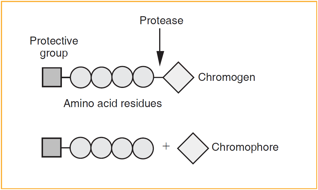
Their method was in principle the same as described above, except that residual factor Xa is measured by using a synthetic factor Xa chromogenic substrate. In addition to this two-stage assay also chromogenic one-stage assays have been introduced. The chromogenic methods enables more precise determination of both UF heparins and LMW heparins, especially given that the methods have also been successfully automated.
Peptide substrates used in the chromogenic anti-Xa assay are generally composed of 3-4 amino acids, with the chromogenic group para-nitroaniline (pNA) attached to the end. When the synthetic substrate is incubated with factor Xa it is cleaved and a chromophore (yellow color) is liberated. This is measured at 405 nm, either during the reaction (kinetic method), or after stopping the reaction with acetic or citric acid (end-point method). The resulting photometric signal is inversely proportional to the heparin activity in the sample.
Chromogenic anti-FXa assays are performed either as single-stage or as two-stage assays. In the latter, exogenous antithrombin is added to the test and the results are then considered to reflect the total heparin concentration. The single-stage assay utilizes only the endogenous antithrombin in the plasma sample, yielding results which are referred to as the effective heparin concentration. The effect of adding antithrombin to the test is apparent in experiments utilizing heparin spiked samples. If the antithrombin activity in the plasma is below the normal range, a decreased heparin recovery is obtained, unless exogenuos antithrombin is added to the test.
However, when plasma samples from heparinized patients with antithrombin levels varying from 35% to 130% were tested with the single-stage method (Chromogenix Coamatic® Heparin) addition of exogenous antithrombin had no effect on the resulting heparin level. Consequently, this kit method is insensitive to variation in the endogenous antithrombin activity in the plasma.
Chromogenic substrates for factor Xa
- Chromogenix S-2222™ Bz-Ile-Glu-(γ-OR´)-Gly-Arg-pNA
- Chromogenix S-2765™ Z-D-Arg-Gly-Arg-pNA
- Chromogenix S-2772™ Ac-D-Arg-Gly-Arg-pNA
- Chromogenix S-2732™ Suc-Ile-Glu-(γ-Piperidyl)-Gly-Arg-pNA
Abbreviations: Bz; benzoyl, Z; benzyloxycarbonyl, R’= H (50%) and R’= CH3 (50%), pNA; 4-nitroaniline
Heparin Monitoring Products
- Chromogenix Coamatic® Heparin
- Chromogenix Coatest® Heparin